Retatrutide mechanism of action: what starts the changes?
Hunger can ease before you know what changed. Researchers made retatrutide from joined protein parts. It copies GIP, GLP-1 and glucagon, hormones carrying body messages. Your hunger, insulin release and use of stored fuel can change.
What is the short answer about changes inside your body?
You may feel less hungry without noticing changes inside cells. Retatrutide attaches to proteins at each cell's surface. Those proteins pass messages inward and change what cells do.
You can't feel a single cell receiving a message.
Some cells release insulin; others help your body burn fuel. Messages reaching the brain can change your interest in food. Researchers studied both cell actions and changes in people.
Cell findings can't settle your benefits and risks.
How does retatrutide work follows the whole body's changes. Here you'll read what starts those actions. Retatrutide hasn't received FDA approval as prescription medicine.
Your treatment needs more than an explanation from a lab.
Why does retatrutide remain in blood after a study shot?
Drug can remain in blood after you stop feeling a shot. Researchers made retatrutide, also called LY3437943, in a lab. They adapted GIP, a gut hormone that helps release insulin.
Retatrutide isn't a hormone your body makes naturally.
Its lab weight is 4731 daltons. Daltons measure tiny particles, rather than your body weight. That weight doesn't tell you how long drug remains in blood. An added fatty part helps explain the slower removal.
You don't have a shot amount from that weight.
The fatty part joins retatrutide to a protein in blood. That larger protein carries the drug through the blood. Your kidneys remove the attached drug more slowly. Drug levels drop halfway over about 6 days [4].
That helped researchers plan their weekly shots.
Retatrutide was also made to copy GLP-1 and glucagon. The gut hormone eases hunger and helps control sugar. Glucagon helps your liver release sugar and use fuel.
Lab images showed attachment at all three hormone sites [3].
You still need patient findings to judge the drug.
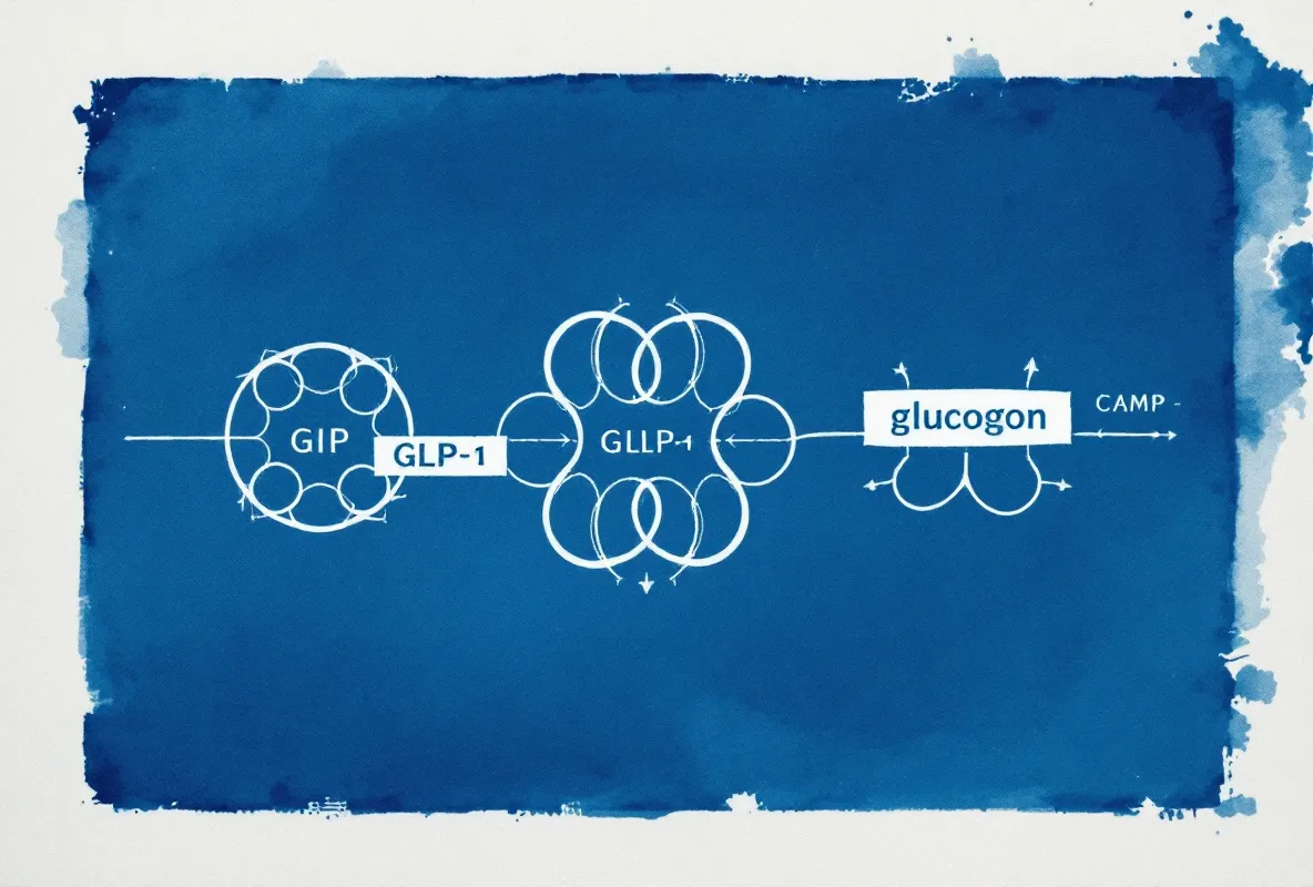
How can a message to a cell change sugar or hunger?
Your insulin helps sugar leave blood for use in cells. Retatrutide can help cells release more insulin. The change begins when the drug attaches outside a cell.
You can't feel each cell responding.
Hormones use proteins built into the cell's outer covering. Part of each protein faces outside; another part faces inside. Drug attachment starts changes on the inside too.
Researchers examined proteins passing those messages through cells [3].
What happens next depends on the kind of cell. Insulin-making cells can respond by releasing insulin. Messages in parts of your brain can reduce interest in food.
Your appetite doesn't measure how much insulin changed.
GIP helps bring out insulin and affects fat use. Glucagon acts on the liver, where sugar is stored. When sugar is too low, glucagon helps release that sugar. The liver can also make sugar from other fuels.
Glucagon helps burn fat; some fat tissue makes heat [1][3][10].
Retatrutide combines sugar-lowering actions with glucagon's sugar-raising action. Researchers checked the combined result in people. People with diabetes had better sugar control.
You can't judge the whole drug from one cell's action.
Your doctor needs patient findings, along with your health history. The lab explanation doesn't settle treatment benefits or lasting safety.
What could microscope images tell you about drug effects?
Your weight-loss questions led researchers to examine tiny drug attachments. A 2024 Cell Discovery paper reported the lab tests [3]. Researchers quickly froze proteins attached to retatrutide. They used thousands of microscope images to study the shapes.
Those images didn't measure your symptoms.
One attachment site normally responds to GLP-1 [3]. That's the gut hormone that affects insulin and hunger. Another site responds to GIP, which also helps release insulin [3]. The third site responds to glucagon, involved in liver sugar and fuel [3].
Retatrutide attached at all three sites.
Researchers called one gripping part loop 1 [3]. That part helps hold the drug against the cell protein. The shape differed among the attachment sites.
Your hunger can't be predicted from a protein's shape.
The shapes may help explain why the drug's hormone actions differ. Researchers also tested cells kept in a dish. Retatrutide started changes inside those cells [3]. The tests explored drug actions without measuring patients' sugar or weight.
You don't have a predicted health benefit from those cell tests.
You can't judge benefits without knowing what happened to people. Patient studies must also check stomach trouble and heart effects. The microscope study can't settle those concerns for you.
Did the combined hormone effects push sugar too high?
Your liver can release sugar while also burning stored fat. Glucagon helps both actions, so researchers watched for high sugar. They wanted more fuel use without an unhealthy sugar rise.
You can't judge that balance without blood tests in people.
Phase 2 means checking benefits and risks in larger groups. Those tests found weight loss without a meaningful rise in fasting sugar [1]. Fasting means the person hadn't eaten before the blood test. The result reflects all the drug's hormone actions together.
One sugar-raising action doesn't predict the whole drug's result.
Retatrutide also copies GIP, a hormone from your gut. GIP helps release insulin, which lowers sugar in your blood. A 2024 Cell review discussed combining GIP with hunger reduction [10]. The authors proposed added effects on fat use and food's appeal.
Those ideas may help explain greater weight losses.
Researchers saw differences in drug attachment during lab work [3]. But attachment alone can't tell you how much each action matters. Your body's response needs evidence from people.
You can't forecast your loss from lab images.
A 2025 paper measured blood changes in people [14]. Blood fats fell alongside proteins that affect fat removal. Researchers used those measurements to study glucagon's role.
Lower protein levels alone aren't proof of better health.
The blood tests don't establish lasting heart protection for you.
Why might food occupy less of your thoughts?
Your brain helps decide when food feels appealing. GLP-1, a gut hormone, also acts in the brain [12]. Brain areas involved in hunger and fullness respond to the hormone. Changed messages can reduce people's interest in seeking food.
Researchers think that helps explain appetite changes.
People trying research products outside trials also report fewer food thoughts. Those accounts weren't checked against a comparison group. They aren't controlled-study findings. Retatrutide trials haven't confirmed those users' reported experiences.
You can't infer a proved brain benefit from their accounts.
A 2025 Medical Sciences review discussed GLP-1 and craving [12]. Craving means a strong urge for something, such as food. The review examined brain changes that might reduce food's appeal.
That explanation doesn't predict your own weight loss.
GIP helps release insulin; glucagon helps your liver use stored fuel. A report about appetite can't separate those body changes. Feeling less hungry doesn't measure what changed inside your brain.
Your mood could change for other reasons too.
Some outside users describe brighter moods and less worry around food. Those accounts may fit known gut-hormone effects in the brain. Studies comparing groups haven't proved those retatrutide benefits.
Your own experience can't be predicted from theirs.
What remains unknown about your heart, kidneys and strength?
Your strength matters when getting out of a chair. Heart and kidney health matter alongside lost weight. Brief studies can't settle safety over years. Larger, longer retatrutide tests continue checking those questions.
Your scales don't give you a complete health answer.
What could a faster pulse mean? Larger study amounts raised pulses near 5-7 beats per minute [1]. Glucagon affects cells setting your heart's pace. The same hormone also acts on the liver's fuel use.
Fuel burning doesn't establish protection for your heart.
Another study is checking lasting heart benefit or harm. You can't settle those effects from pulse readings alone.
What do kidney studies check? TRANSCEND-CKD examines how kidneys respond to retatrutide. Researchers measure how well the kidneys filter blood. They also check urine for protein leaking through damaged kidneys.
Those tests address concerns you can't judge by weighing yourself.
Some approved drugs that ease hunger have shown kidney protection. Researchers don't yet know how added glucagon changes that benefit. Another drug's findings can't promise the same protection for you.
You won't settle your kidney question with another drug's findings.
Could muscle loss make standing harder? A 2025 Lancet Diabetes & Endocrinology paper examined lost tissue besides fat. Muscle was included in those measurements. Caution 5 on the effects page explains why muscle matters.
Your legs need muscle to lift and carry you.
Eating much less may explain some muscle loss. Direct drug effects might also play a part. Researchers haven't settled those possible causes.
You can't judge strength from the weight-loss figures alone.